
Technology
How does a picosecond laser differ from a Q-switched laser?
Picosecond and Q-switched lasers break up tattoo ink and pigment with pulses lasting picoseconds or nanoseconds, so short that the energy stays in the particle until it fragments. We supply three devices and one handpiece in this class: PicoSure Pro, a 755 nm alexandrite laser that also works at 532 and 1064 nm with optional handpieces; PicoPlus, an Nd:YAG laser with picosecond pulses and a nanosecond mode, at four wavelengths: 1064, 532, 660 and 595 nm; Hollywood Spectra, a Q-switched Nd:YAG laser with additional handpieces; and the handPICO handpiece for Etherea MX, with picosecond pulses at 1064 and 532 nm.
Translated from Dutch with the help of AI tools.
Why ink needs a short pulse
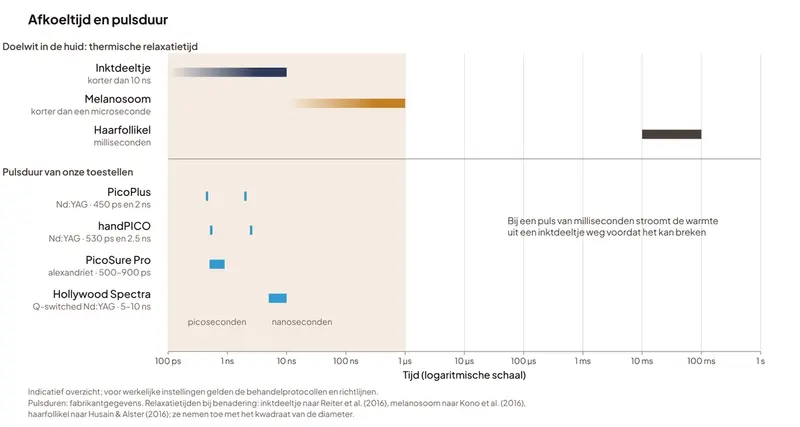
A laser only targets pigment selectively when two conditions are met. The pigment must absorb the wavelength more strongly than the surrounding tissue, and the pulse must be shorter than its thermal relaxation time: the time it needs to lose its heat [1].
Thermal relaxation time depends on size. A hair follicle takes milliseconds to cool; an ink particle or pigment granule is much smaller and cools within a microsecond or less [1][2][3].
For ink, a millisecond pulse is therefore too long: the heat leaks away before the particle gets hot enough to fragment. That is why tattoo ink and dermal pigment are treated with lasers that deliver nanosecond or picosecond pulses. Superficial pigmented lesions are the exception: they also respond to 532 nm with millisecond pulses and to IPL.
Nanoseconds: Q-switching
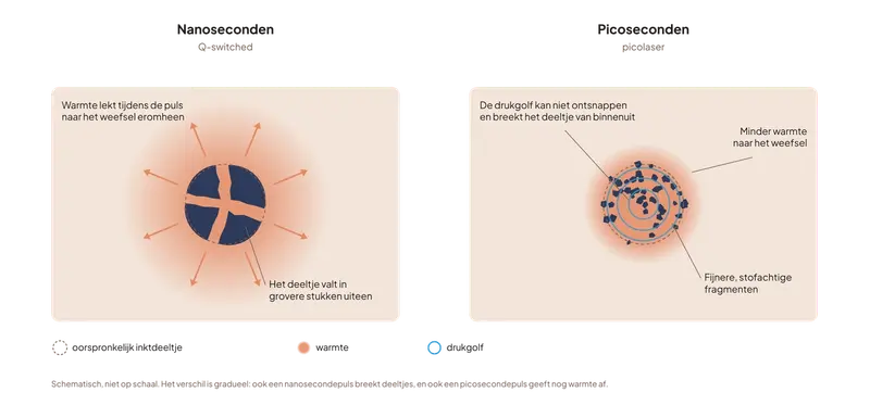
A Q-switched laser releases its energy in pulses of a few nanoseconds. That is shorter than the thermal relaxation time of an ink particle, so the heat is confined to the particle, which heats so rapidly that it fragments. A mechanical effect contributes as well as the heat [4][5].
Hollywood Spectra works this way, with pulses of 5 to 10 nanoseconds. PicoPlus and handPICO have a nanosecond mode as well as their picosecond pulses, at 2 and 2.5 nanoseconds respectively.
Picoseconds: the pressure stays in the particle
A picosecond pulse lasts less than a nanosecond. When a particle absorbs light, it expands and a pressure wave travels through it. If the pulse is shorter than the time that wave needs to cross the particle, the pressure cannot escape and fractures the particle from within. This is known as stress confinement [5].
The photothermal component is therefore smaller than with nanosecond pulses, and the tissue around the particle receives less heat [5]. The ink breaks into finer, dust-like particles. The difference from Q-switching is one of degree: a nanosecond pulse also breaks particles, and a picosecond pulse still releases some heat. PicoPlus delivers pulses of 450 picoseconds, handPICO of 530 picoseconds and PicoSure Pro of 500 to 900 picoseconds.
How the fragments are cleared
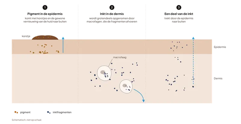
After the pulse, the ink is broken up but not yet gone: the body has to clear the fragments. Epidermal pigment is shed with crusting and normal epidermal turnover. Ink in the dermis is largely taken up by macrophages, which carry the fragments away [4].
Some of the ink is also eliminated through the epidermis: after a picosecond treatment, ink particles have been observed migrating outwards through all layers of the epidermis [6].
Which wavelength targets which colour
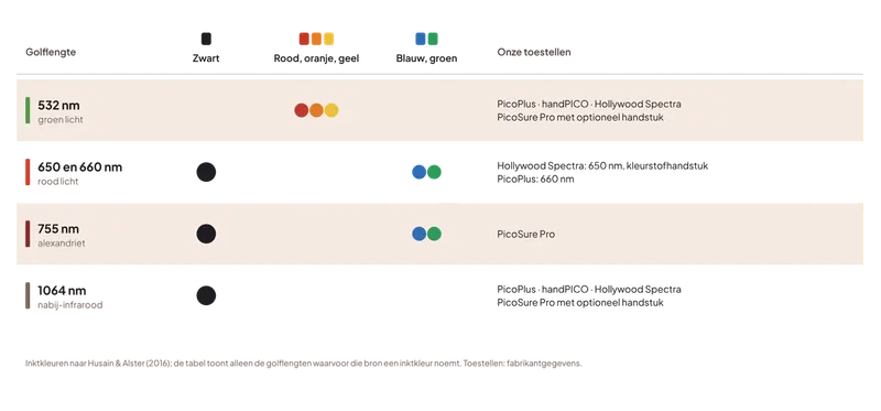
Which ink colours respond depends on the wavelength. Black ink absorbs across the red and near-infrared range. Red, orange and yellow absorb mainly green light, so they need 532 nm. Blue and green ink absorb between 600 and 800 nm, a range that includes 755 nm and the 650 and 660 nm handpieces [7]. In a series of twelve tattoos with blue or green pigment, at least 75% of the blue and green had cleared after one or two treatments with a 755 nm picosecond alexandrite laser [8].
The same principle applies to melanin. Green 532 nm light is strongly absorbed and penetrates only superficially, which suits it to epidermal pigment such as solar lentigines. At 1064 nm, melanin absorbs less and the light reaches the deeper dermis, the site of dermal pigment such as naevus of Ota. Because the epidermis absorbs little of it, 1064 nm is also used in darker skin types [7][9].
Our devices by wavelength:
- PicoSure Pro: 755 nm, with optional handpieces that convert that light to 532 or 1064 nm.
- PicoPlus: 1064 and 532 nm, with handpieces for 595 and 660 nm.
- handPICO: 1064 and 532 nm.
- Hollywood Spectra: 1064 and 532 nm, with additional handpieces for 585 and 650 nm.
Fractional picosecond treatment
The same picosecond pulse can also be used for indications other than pigment. A fractional handpiece splits the beam into a grid of small columns in which the energy density is much higher than in the rest of the beam. In a histological study with the fractional lens of a picosecond alexandrite laser, this produced microscopic cavities in the epidermis. The author attributes them to laser-induced optical breakdown (LIOB), a localised plasma that is triggered when melanin absorbs the light. These zones were shed within about three weeks, and in the dermis the author describes new collagen and elastic tissue [10].
On PicoSure Pro this lens is called the Focus™ Lens Array; handPICO has an optional fractional microlens array at 1064 nm. These handpieces are mainly used for (acne) scars and skin rejuvenation.
What it is used for
Tattoo removal
Tattoo ink fragments only with nanosecond or picosecond pulses; with a longer pulse, the heat diffuses out of the ink particle [1]. Both regimes remove ink, but picosecond lasers often need fewer sessions [11][12]. Even recalcitrant tattoos that no longer fade after Q-switched treatment can still respond well to a picosecond laser [5]. Which ink colour responds depends on the wavelength [7].
Permanent makeup removal
Permanent make-up (PMU) pigment is broken up with nanosecond or picosecond pulses, with the wavelength chosen to match the colour of the pigment [7]. Skin-coloured, white and rust-coloured PMU pigment often contains iron oxide or titanium dioxide, which can turn black under such a short pulse (paradoxical darkening) [7].
Pigmentation
Dermal pigment, such as naevus of Ota, needs nanosecond or picosecond pulses; superficial spots such as lentigines and freckles also respond to 532 nm with millisecond pulses and to IPL [3][13][14]. For naevus of Ota, picosecond alexandrite gave better clearance than Q-switched alexandrite in a randomised study [15]. In Asian skin, picosecond lasers caused less pigmentary change than Q-switched lasers in comparative studies [16].
Melasma
For melasma, Q-switched and picosecond lasers at 1064 nm are used for laser toning: a series of low-energy treatments, as an adjunct to topical treatment rather than a replacement for it [17][18]. Most of the evidence concerns Q-switched toning [19]. Picosecond lasers gave a comparable result in two randomised split-face studies, one of them with less pain after the first session [20][21]. Melasma often recurs, and mottled hypopigmentation has been reported after many sessions with a high cumulative energy [17].
(Acne) scars
With a fractional picosecond handpiece at 1064 nm, acne scars improved in a randomised split-face study comparably to a 1550 nm non-ablative fractional laser, with less pain but more pinpoint bleeding [22]. Compared with fractional CO₂, physicians in another split-face study rated the CO₂ side as more improved, while the picosecond side had fewer side effects [23]. A meta-analysis found less post-inflammatory hyperpigmentation than with other fractional lasers [24].
Skin quality improvement
At 1064 nm, pores improved more with a fractional picosecond handpiece (microlens) than with quasi-long pulses in a split-face study of 25 Asian patients [25].
Skin rejuvenation
A fractional picosecond handpiece concentrates the energy into a grid of small columns. With the fractional lens of a picosecond alexandrite laser, a histological study describes small zones in the epidermis, followed by new collagen and elastic tissue in the dermis [10]. A systematic review lists skin rejuvenation among the clinically supported applications of picosecond lasers [26].
Picosecond versus Q-switched
Finer fragmentation with less heat does not translate into a better result for every indication; the comparison has been studied most extensively in tattoos.
Tattoos
As early as 1998, a study compared two laboratory lasers on the same black tattoos: in twelve of sixteen, the half treated with 35-picosecond pulses faded more than the half treated with 10-nanosecond pulses [27]. A 2026 systematic review of 46 studies on Nd:YAG lasers found that picosecond lasers sometimes reached comparable clearance with fewer sessions or less energy, without a consistent advantage over Q-switched. Black and blue ink usually needed four to eight sessions for at least 75% clearance [11]. In a French randomised study of 49 patients, mostly with black and blue professional tattoos, 33% of tattoos reached that level of clearance with the picosecond laser, versus 14% with the nanosecond laser [12].
Pigment
A meta-analysis of twenty randomised studies found comparable clearance of pigmentary disorders with picosecond and nanosecond lasers. Picosecond lasers caused less pain and less post-inflammatory hyper- and hypopigmentation, a difference shown mainly in Asian patients [16].
For solar lentigines, the 532 nm picosecond laser gave greater clearance after one treatment in a randomised split-face study of twenty patients, with post-inflammatory hyperpigmentation in 5% versus 30% after Q-switched treatment [28]. Another randomised study, in Asian patients, found no difference in clearance, although patients were more satisfied with the picosecond side [29].
For naevus of Ota, picosecond alexandrite gave better clearance than Q-switched alexandrite in a randomised split-lesion study of 56 patients, with fewer sessions, less pain and less hypo- and hyperpigmentation [15].
Melasma
For melasma, two randomised split-face studies at 1064 nm found no difference in efficacy between picosecond and Q-switched toning, and the more recent one found no difference in recurrence either; in that study, pain after the first session was lower with picosecond [20][21]. A systematic review of 42 studies describes low-energy Q-switched toning as generally effective and safe, with the recognised drawback that melasma can relapse within a few months [17]. An international consensus places lasers for melasma after topical agents and oral tranexamic acid, and recommends using them with great caution to avoid worsening the condition [18].
Skin type and safety
Whichever pulse regime is used, skin type helps determine the risk of pigmentary change. The more melanin the epidermis contains, the more light it absorbs itself. For darker skin types, review articles therefore favour 1064 nm and regard 532 nm as higher risk [7][9]. In a Korean multicentre study, 20% of patients developed post-inflammatory hyperpigmentation after Q-switched 532 nm for solar lentigines [30].
- Tattoos and permanent make-up. Permanent depigmentation has been reported after many sessions with Q-switched lasers [9]. Skin-coloured, white and rust-coloured pigment can turn black [7].
- Laser toning for melasma. Mottled hypopigmentation can develop after repeated sessions, especially with a high cumulative energy. Used too aggressively, toning can also cause hyperpigmentation, especially in dark skin [17].
Laser light from these devices can damage the retina; patient and operator wear protective eyewear rated for the wavelength in use. A pigmented lesion is only treated with a laser once a benign diagnosis has been established.
When another technology is the better choice
For some pigmentary conditions, another technology is more appropriate than a picosecond or Q-switched laser.
Long-pulse alexandrite and Nd:YAG
With millisecond pulses, the same laser crystals heat hair follicles and blood vessels, but they have little effect on dermal pigment and tattoo ink, because the heat flows out of the particle [1][3]. See our alexandrite and Nd:YAG pages.
Vascular lasers and IPL
Superficial pigmented lesions, such as solar lentigines and freckles, also respond to long-pulse 532 nm [13][14] and to IPL [3]. In Chinese patients, long-pulse 532 nm caused no more pigmentary change than Q-switched 532 nm [13][14]. Dermal pigment still needs nanosecond or picosecond lasers [3].
Advice
Is this technology right for your practice?
A single page cannot fully cover the choice between technologies. It depends on your patients and their skin types, the indications you want to treat, the recovery time your patients will accept and the equipment you already have. Our clinical and sales specialists have years of experience with that choice and are glad to help you make it.
Already working with one of our devices? You will find the treatment protocols and guidelines on the customer portal, and we go deeper into technique in the training after delivery and at our Academy events.
This is an indicative overview to explain how technologies differ. Actual settings are governed by the treatment protocols and guidelines.
References
- Anderson RR, Parrish JA. Selective photothermolysis: precise microsurgery by selective absorption of pulsed radiation. Science. 1983;220(4596):524–527. doi:10.1126/science.6836297
- Reiter O, Atzmony L, Akerman L, Levi A, Kershenovich R, Lapidoth M, Mimouni D. Picosecond lasers for tattoo removal: a systematic review. Lasers Med Sci. 2016;31(7):1397–1405. doi:10.1007/s10103-016-2001-0
- Kono T, Shek SY, Chan HH, Groff WF, Imagawa K, Akamatsu T. Theoretical review of the treatment of pigmented lesions in Asian skin. Laser Ther. 2016;25(3):179–184. doi:10.5978/islsm.16-or-13
- Barua S. Laser-tissue interaction in tattoo removal by Q-switched lasers. J Cutan Aesthet Surg. 2015;8(1):5–8. doi:10.4103/0974-2077.155063
- Kasai K. Picosecond laser treatment for tattoos and benign cutaneous pigmented lesions (secondary publication). Laser Ther. 2017;26(4):274–281. doi:10.5978/islsm.17-RE-02
- Nguyen L, Mess C, Huck V, Schneider SW, Herberger K. Intravital visualization of tattoo particles after picosecond laser treatment. Skin Res Technol. 2025;31(10):e70283. doi:10.1111/srt.70283Carried out with PicoPlus; nine participants, eleven tattoos, one treatment, imaging at 6 and 12 weeks. Two authors received speaker fees from Cynosure Lutronic.
- Husain Z, Alster TS. The role of lasers and intense pulsed light technology in dermatology. Clin Cosmet Investig Dermatol. 2016;9:29–40. doi:10.2147/CCID.S69106
- Brauer JA, Reddy KK, Anolik R, Weiss ET, Karen JK, Hale EK, Brightman LA, Bernstein L, Geronemus RG. Successful and rapid treatment of blue and green tattoo pigment with a novel picosecond laser. Arch Dermatol. 2012;148(7):820–823. doi:10.1001/archdermatol.2012.901Twelve tattoos, no control group.
- Harnchoowong S, Vachiramon V, Jurairattanaporn N. Cosmetic considerations in dark-skinned patients. Clin Cosmet Investig Dermatol. 2024;17:259–277. doi:10.2147/CCID.S450081
- Tanghetti EA. The histology of skin treated with a picosecond alexandrite laser and a fractional lens array. Lasers Surg Med. 2016;48(7):646–652. doi:10.1002/lsm.22540
- Wong JJE, Khan S, Tandel M, Dajani Z, Seth I. Efficacy of Nd:YAG lasers for tattoo removal: systematic review of clinical outcomes, clearance rates, and treatment parameters. J Cosmet Dermatol. 2026;25(6):e70914. doi:10.1111/jocd.70914
- Lorgeou A, Perrillat Y, Gral N, Lagrange S, Lacour JP, Passeron T. Comparison of two picosecond lasers to a nanosecond laser for treating tattoos: a prospective randomized study on 49 patients. J Eur Acad Dermatol Venereol. 2018;32(2):265–270. doi:10.1111/jdv.14492
- Chan HH, Fung WK, Ying SY, Kono T. An in vivo trial comparing the use of different types of 532 nm Nd:YAG lasers in the treatment of facial lentigines in Oriental patients. Dermatol Surg. 2000;26(8):743–749. doi:10.1046/j.1524-4725.2000.00039.x
- Ho SG, Chan NP, Yeung CK, Shek SY, Kono T, Chan HH. A retrospective analysis of the management of freckles and lentigines using four different pigment lasers on Asian skin. J Cosmet Laser Ther. 2012;14(2):74–80. doi:10.3109/14764172.2012.670707Retrospective, 40 patients divided across four lasers.
- Ge Y, Yang Y, Guo L, Zhang M, Wu Q, Zeng R, Rong H, Jia G, Shi H, Fang J, Lin T. Comparison of a picosecond alexandrite laser versus a Q-switched alexandrite laser for the treatment of nevus of Ota: a randomized, split-lesion, controlled trial. J Am Acad Dermatol. 2020;83(2):397–403. doi:10.1016/j.jaad.2019.03.016
- Wu W, Du Y, Zhang Y, Su Q, Wang F. Comparative appraisal with meta-analysis of picosecond versus nanosecond lasers for hyperpigmented disorders and tattoos. Lasers Med Sci. 2025;40(1):465. doi:10.1007/s10103-025-04721-3
- Lee YS, Lee YJ, Lee JM, Han TY, Lee JH, Choi JE. The low-fluence Q-switched Nd:YAG laser treatment for melasma: a systematic review. Medicina (Kaunas). 2022;58(7):936. doi:10.3390/medicina58070936
- Passeron T, Desai SR, Abdallah M, Al-Niaimi F, Dlova N, Grimes PE, Ocampo-Candiani J, Sarkar R, Xiang LF, Miot HA. Global consensus on the management of melanin hyperpigmentation disorders. J Eur Acad Dermatol Venereol. 2026;40(5):760–772. doi:10.1111/jdv.70185
- Chehrara M, Tabavar A, Roohaninasab M, Jafary P, Mohammad AP, Mozafarpoor S, Alavi Rad E, Goodarzi A. The efficacy of laser therapy in melasma treatment: a systematic review and meta-analysis. J Cosmet Dermatol. 2025;24(12):e70602. doi:10.1111/jocd.70602
- Hong JK, Shin SH, Park SJ, Seo SJ, Park KY. A prospective, split-face study comparing 1,064-nm picosecond Nd:YAG laser toning with 1,064-nm Q-switched Nd:YAG laser toning in the treatment of melasma. J Dermatolog Treat. 2022;33(5):2547–2553. doi:10.1080/09546634.2022.2033674
- Wu Y, Wang Q, Feng J, Chen Z, Huang L, Feng Y, Li X. Comparison of 1064-nm picosecond and Q-switched neodymium-doped yttrium aluminum garnet lasers for melasma in Asian women: a randomized double-blind split-face trial. J Cosmet Dermatol. 2026;25(7):e71008. doi:10.1111/jocd.71008
- Chayavichitsilp P, Limtong P, Triyangkulsri K, Pratumchart N. Comparison of fractional neodymium-doped yttrium aluminum garnet (Nd:YAG) 1064-nm picosecond laser and fractional 1550-nm erbium fiber laser in facial acne scar treatment. Lasers Med Sci. 2020;35(3):695–700. doi:10.1007/s10103-019-02891-5
- Yuan Y, He Y, Fang J, Zhang M, Wu Q. Comparison of the fractionated Nd:YAG 1064-nm picosecond laser with holographic optics and the fractional CO₂ laser in atrophic acne scar treatment: a prospective, randomized, split-face study. Int J Dermatol. 2025;64(1):85–91. doi:10.1111/ijd.17296
- Li J, Duan F, Kuang J. Fractional picosecond laser for atrophic acne scars: a meta-analysis. J Cosmet Dermatol. 2023;22(8):2205–2217. doi:10.1111/jocd.15862
- Yim S, Lee YH, Choi YJ, Kim WS. Split-face comparison of the picosecond 1064-nm Nd:YAG laser using a microlens array and the quasi-long-pulsed 1064-nm Nd:YAG laser for treatment of photoaging facial wrinkles and pores in Asians. Lasers Med Sci. 2020;35(4):949–956. doi:10.1007/s10103-019-02906-1
- Wu DC, Goldman MP, Wat H, Chan HHL. A systematic review of picosecond laser in dermatology: evidence and recommendations. Lasers Surg Med. 2021;53(1):9–49. doi:10.1002/lsm.23244
- Ross V, Naseef G, Lin G, Kelly M, Michaud N, Flotte TJ, Raythen J, Anderson RR. Comparison of responses of tattoos to picosecond and nanosecond Q-switched neodymium:YAG lasers. Arch Dermatol. 1998;134(2):167–171. doi:10.1001/archderm.134.2.167Two laboratory lasers, not commercial devices.
- Kim JY, Yang J, Huh G, Choi YJ, Kim WS. A split-face, single-blinded, randomized controlled comparison of 532 nm picosecond neodymium-doped yttrium aluminum garnet laser versus 532 nm Q-switched neodymium-doped yttrium aluminum garnet laser in the treatment of solar lentigines. Ann Dermatol. 2020;32(1):8–13. doi:10.5021/ad.2020.32.1.8
- Vachiramon V, Iamsumang W, Triyangkulsri K. Q-switched double frequency Nd:YAG 532-nm nanosecond laser vs. double frequency Nd:YAG 532-nm picosecond laser for the treatment of solar lentigines in Asians. Lasers Med Sci. 2018;33(9):1941–1947. doi:10.1007/s10103-018-2560-3
- Kang HJ, Na JI, Lee JH, Roh MR, Ko JY, Chang SE. Postinflammatory hyperpigmentation associated with treatment of solar lentigines using a Q-switched 532-nm Nd:YAG laser: a multicenter survey. J Dermatolog Treat. 2017;28(5):447–451. doi:10.1080/09546634.2016.1254330